Percolation of Ion-Irradiation-Induced Disorder in Complex Oxide Interfaces
Abstract
Mastery of order-disorder processes in highly non-equilibrium nanostructured oxides has significant implications for the development of emerging energy technologies. However, we are presently limited in our ability to quantify and harness these processes at high spatial, chemical, and temporal resolution, particularly in extreme environments. Here we describe the percolation of disorder at the model oxide interface LaMnO3 / SrTiO3, which we visualize during in situ ion irradiation in the transmission electron microscope. We observe the formation of a network of disorder during the initial stages of ion irradiation and track the global progression of the system to full disorder. We couple these measurements with detailed structural and chemical probes, examining possible underlying defect mechanisms responsible for this unique percolative behavior.
I Introduction
Interface engineering of oxide thin films is one of the great achievements of materials science, underpinning exotic physicsHwang et al. (2012) and giving rise to advanced computing and energy technologies.Martin and Rappe (2017) Because of strong interplay among lattice, charge, and spin degrees of freedom, even slight fluctuations in local order at oxide interfaces can greatly impact behaviors such as electronicHuang et al. (2018) and ionic conductivity.Fabbri, Pergolesi, and Traversa (2010) Order can be disrupted via numerous defect mechanisms, such as local structural perturbationsIsmail-Beigi et al. (2017); Shamblin et al. (2016a) or the formation of cation and anion vacancies.Gunkel et al. (2020); Zhang et al. (2015) The desire to understand these mechanisms has motivated efforts to map defect formation pathways during oxide synthesisMacManus-Driscoll et al. (2020); Brahlek et al. (2018) and exposure to extreme environments.Spurgeon (2020); Zhang et al. (2018); Beyerlein et al. (2013) Understanding oxides in extremes of temperature and irradiation is particularly important, since devices such as solid oxide fuel cells (SOFCs),Shamblin et al. (2016a); Tuller and Bishop (2011) durable nuclear waste forms,Ewing, Weber, and Lian (2004); Sickafus et al. (2000) and space-based electronicsCramer et al. (2016) must deliver reliable long-term performance under challenging conditions. At a more fundamental level, the nature of order-disorder processes represents a grand scientific question, with implications for the design of high entropy alloysGeorge, Raabe, and Ritchie (2019) and ceramics,Oses, Toher, and Curtarolo (2020) strongly correlated quantum systems,Vepsäläinen et al. (2020) and more.
To dictate order-disorder behavior, we must be able to visualize and direct defect formation processes at high spatial, chemical, and temporal resolution. The community has a long, successful history of using tailored ion irradiation to manipulate and induce defect populations in bulk metalsWas (2015); Odette, Alinger, and Wirth (2008) and oxides.Meldrum, Boatner, and Ewing (1998); Lumpkin et al. (2009); Sickafus et al. (2000); Shamblin et al. (2016b); Lang et al. (2010) While recent studiesZhang et al. (2018); Martínez, Uberuaga, and Beyerlein (2016); Beyerlein et al. (2015, 2013) have showcased the promising properties of nanostructured systems, such as enhanced defect annihilation and radiation hardness, past work has focused almost exclusively on metals and bulk oxides.Zhang et al. (2018) A handful of studies of model oxide interfaces have combined controlled ion irradiation and local characterization tools to identify unique behaviors such as anti-site defect buildup,Kreller et al. (2019) oxygen vacancy formation,Spurgeon et al. (2020) and orientation-/chemistry-dependent amorphization behavior.Kaspar et al. (2017); Aguiar et al. (2014a, b) These studies show that there is an interplay between defect formation energy, which depends on the interface configuration (e.g. strain, charge state, chemistry)Spurgeon et al. (2020); Dholabhai et al. (2014); Aguiar et al. (2014a, b); Zhuo et al. (2011) and defect kinetics, particularly defect mobility in different interface components.Zhuo et al. (2012) However, most prior work has focused on static snapshots of these materials and has not possessed sufficient spatiotemporal resolution to probe the kinetics of local defect formation, particularly during the initial loss of crystallinity in oxide thin films.
Moving beyond traditional static characterization approaches toward in situ methods will allow us to capture the initial onset and evolution of local defects. In situ (scanning) transmission electron microscopy ((S)TEM) has been widely employed by the radiation effects community to examine local defects in materials,Parrish et al. (2021); Lian et al. (2009); Birtcher et al. (2005) providing an invaluable window into irradiation-induced disorder, particularly in the low dose regime. These methods have long been used to examine bulk ceramics,Ye et al. (2011); Lian et al. (2009); Zhang et al. (2005); Wang (1998); Zinkle and Hodgson (1992) revealing radiation-induced defect formation pathways and kinetics. Local (S)TEM probes are particularly well-suited to examining nanostructured oxide interfaces,Spurgeon and Chambers (2018) whose non-equilibrium behavior can deviate greatly from bulk materials, but surprisingly little work has been done on complex oxide thin film systems in the context of radiation damage. More broadly, the microscopy community has also recognized the need for new data-driven approaches to characterization of transient processes, needed to detect and quantify salient features during high-speed imaging.Hattar and Jungjohann (2021); Spurgeon et al. (2021); Taheri et al. (2016)
Here we visualize the evolution of local disorder at a LaMnO3 (LMO) / SrTiO3 (STO) (001) perovskite oxide interface using ion irradiation coupled with in situ high-resolution transmission electron microscopy (HRTEM) imaging at the I3TEM irradiation facility at Sandia National Laboratories. We observe the initial onset of disorder and track its progression over continued irradiation. Using a Fourier filtering time series approach, we show that initial radiation damage is accommodated by the percolation of amorphous regions throughout the crystalline LMO matrix. In addition, we find evidence for a preserved crystalline interface region even at the highest fluence studied, whose origin we examine in the context of electronic structure calculations and past work. Taken together, these results demonstrate the power of high-resolution in situ approaches to derive complex disordering pathways at oxide interfaces. More broadly, the data processing approaches we demonstrate may be applied to other dynamic studies of complex materials phase transitions.
II Results and Discussion
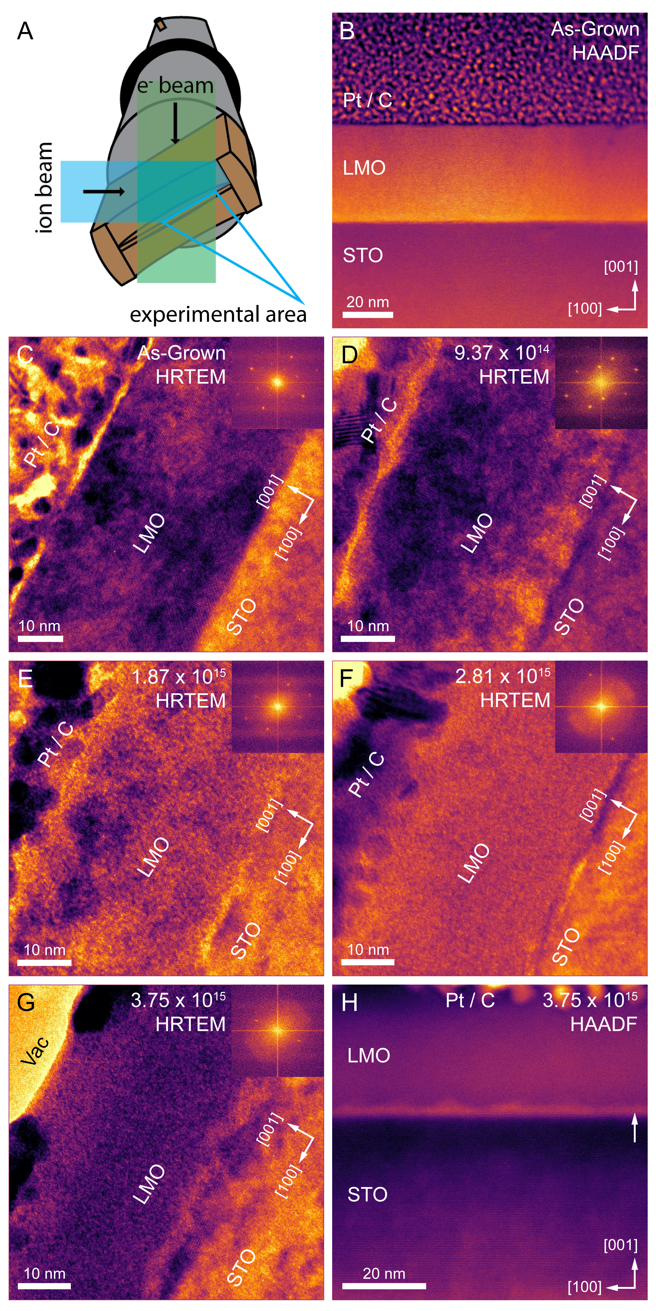
We first consider the overall degradation of the system from the crystalline, as-grown condition to its disordered state. Figure 1.A shows the configuration of the electron and ion beams during irradiation, with the sample inclined 30∘ in the X tilt direction to minimize shadowing of the ion beam. Figure 1.B shows a cross-sectional STEM-HAADF image of the starting epitaxial 40 nm LMO film along the STO [100] crystallographic zone-axis prior to irradiation. In this mode, the directly-interpretable atomic number () contrast reveals a clear difference between the film and substrate, with minimal intermixing, excellent epitaxy, and no extended defects. Figure 1.C shows a HRTEM image of the same film in the I3TEM system, with the interface rotated approximately 60∘ about the [100] zone (the normal direction to the image plane). Phase contrast in HRTEM is less directly interpretable than STEM-HAADF, but the inset fast Fourier transform (FFT) and clear lattice fringes confirm the crystalline starting condition. From this point, a series of four irradiations were performed in 1 hour increments with 2.8 MeV Au4+ ions, as shown in Figures 1.D–G, with the sample inclined during irradiation and then tilted back to the zone for imaging. Each irradiation step corresponds to a fluence of Au4+ cm-2. These figures show a gradual amorphization sequence, starting from the LMO film and extending to the STO substrate. A careful inspection of the images and associated FFTs reveals the emergence of local amorphous patches in the LMO at a fluence of Au4+ cm-2 (Figure 1.D), with less apparent damage in the STO film. By a fluence of Au4+ cm-2 (Figure 1.E), the patches have grown more extensive in the LMO, and the STO side also exhibits obvious disorder; this is accompanied by a decrease in the intensity of the FFT reflections. At a fluence of Au4+ cm-2 (Figure 1.F), the LMO film is almost entirely amorphous, except for a thin, several nm-thick band at the interface. Finally, at a fluence of Au4+ cm-2 (Figure 1.G), the LMO film appears similarly disordered to its state at a fluence of Au4+ cm-2, but it is clearly thinner in the beam direction (as indicated by the larger vacuum region at the top left corner of the image), likely due to sputtering of the film under the ion bombardment. In addition, the STO layer is extensively damaged and the only remaining lattice fringes are from a 2–3 nm band at the film-substrate interface. There is almost no periodic signal left in the FFT, reflecting the loss of overall crystalline order. STEM-HAADF images of the Au4+ cm-2 condition (Figure 1.H) show that some lattice fringes are still present in the bulk of the STO, but the bulk of the LMO is completely amorphous. Nonetheless, there is a pronounced and persistent crystalline band on the film side of the interface (marked by the arrow). We note that SRIM simulations, shown in Supporting Information Note 3, indicate a trace amount of Au ions may be retained in the TEM foil, which may partly influence the kinetics of the radiation response. However, a similar irradiation response has been observed in the La2Ti2O7 / STO system,Spurgeon et al. (2020) where a 5–10 nm crystalline band persisted on the STO side of the interface. While the origins and mechanisms leading to the resistant interface behavior are yet not fully understood, we do note the presence of some cation intermixing in the irradiated sample (shown in Supporting Information Note 4), which may affect its stability. Overall, this points to the broader trend for perovskite oxide interface systems to remain crystalline under irradiation environments.
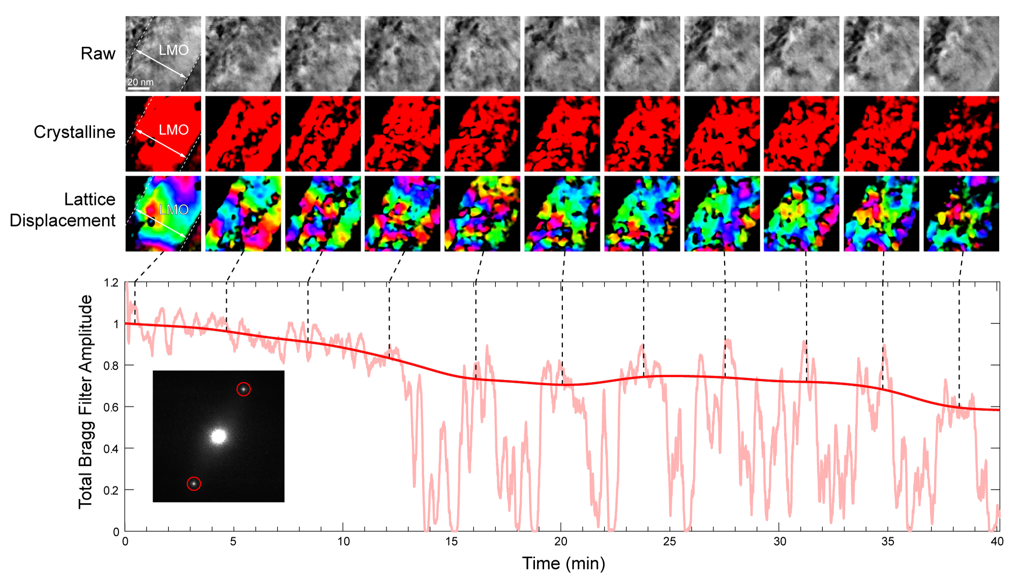
We focus next on the initial stages of the irradiation process, probing the initial percolation of disorder and its buildup to the more global amorphization shown in Figure 1.H. For this experiment, we captured a high-speed time series with the sample inclined at 30∘ in the X tilt direction to maximize exposure to the ion beam. At this high tilt angle it is no longer possible to directly resolve atomic columns in the [100] zone-axis, and we instead image (100)-type lattice planes. However, the remarkable stability of the sample during the extended period of irradiation (40 minutes, total fluence Au4+ cm-2) provided a unique window into the initial stages of disorder. We employ a temporal Fourier filtering approach, analogous to time-resolved geometric phase analysis (GPA),Hÿtch, Snoeck, and Kilaas (1998) processing the raw movie frames into maps of local crystallinity and lattice displacement. In addition, we quantify the total Bragg filter amplitude, which effectively measures the spatial abundance of the selected (100)-type lattice domains in any given frame. It is important to note that while the overall sample was quite stable, these measurements are very challenging and some periodic bending/rotation of the sample due to local heating did occur; since this results in large random drops in the Bragg filter amplitude, a trend line was fit to the Bragg filter amplitude only where crystalline signal is present (see Supporting Information Methods for details). These measurements are shown in Figure 2 and Supporting Information Movie S1.
We see that there is initially a large contiguous block of crystalline region, as expected. By 5 minutes, some disorder emerges in the film, beginning at its center and extending laterally, but the Bragg amplitude is still around 95% of the starting condition. Disorder is accompanied by significant local lattice displacement around the defective regions. At this stage, misfit dislocations emerge that run from the lower left to upper right corner of the frames. Between 9 and 19 minutes, the percolation extends from the film center to its surface and the substrate interface. At this point there is a substantial drop in the Bragg amplitude to nearly 80%, accompanied by a still more complex pattern of lattice rotation and crystalline domains, some nm in size. The misfit dislocations appear to reconnect, forming repaired epitaxial domains, but there is a clear increase in disorder. Between 19 to 33 minutes the overall distribution remains steady near 80%, but between 33 and 37 minutes the percolation continues and the crystalline domains become increasingly sparse and disconnected. In the final condition around 40 minutes, only about 60% of the film is still similar to the starting crystalline condition and there is a large disconnected network of ordered regions. These results point to the highly non-uniform evolution of the interface during the initial stages of irradiation.
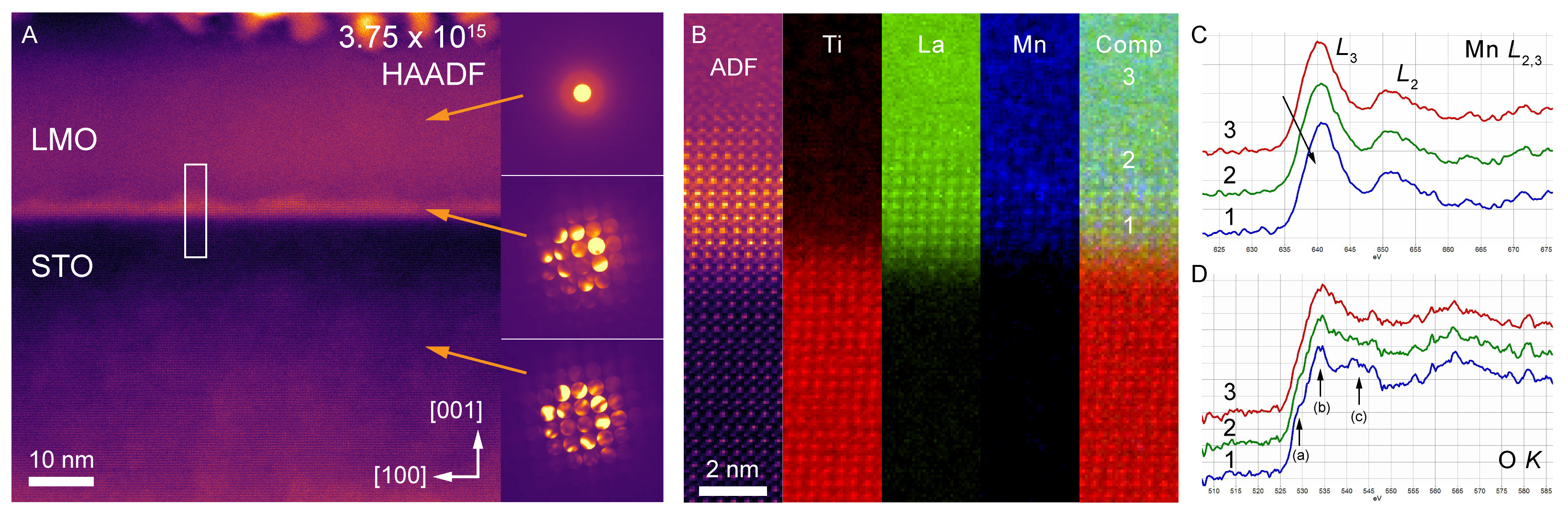
To further explore this non-uniform interface response in later stages of irradiation, we have performed local position-averaged convergent beam electron diffraction (PACBED) and electron energy loss spectroscopy (STEM-EELS) measurements. The former is a mode of scanning nanodiffractionOphus (2019) that can quantify crystallinity at the nanoscale and has been used before to examine local disorder in oxides,Savitzky et al. (2020); Spurgeon et al. (2020); Janish et al. (2019) while the latter has been used to examine chemical states in both pristine and irradiated oxides.Spurgeon (2020) As shown in Figure 3.A, at a fluence of Au4+ cm-2 the bulk of the LMO film has amorphized, resulting in a diffuse, ring-like PACBED pattern. In contrast, PACBED from the uniform nm interface band exhibits strong diffraction disks; there is also evidence for more weakly diffracting patches that extend further into the film. Similarly, the STO side of the interface also exhibits strong diffraction disks, despite some distributed damage, consistent with past observations.Zhang et al. (2005); Jiang et al. (2012) Correlative STEM-EELS chemical analysis of the interface, shown in Figure 3.B, reveals the gradual breakup of the crystalline film upon transitioning from the interface into the bulk, resulting in three distinct regions (labeled 1–3). Region (1) consists of a nm region of LMO that is strongly diffracting and highly chemically ordered on both the La and Mn sublattices. EELS spectra from this region contain a characteristic pre-peak (a), main peak (b), and secondary peak (c) in the O edge fine structure, as well as a clear Mn white-line doublet, consistent with previous observations.Kaspar et al. (2019); Varela et al. (2009) Moving away from the interface and into the LMO, we move into a more weakly diffracting nm Region (2) consisting of poorly defined perovskite atomic order. This region exhibits a reduction in the O pre-peak feature and loss of definition between the main and secondary peaks. In addition, there is a clear 0.5 eV shift of the Mn edge to lower energy loss, pointing toward underlying reduction. This region is followed by third and final amorphous region (3) with no extended ordering of La and Mn; now the O edge pre-peak has disappeared and the main edge features have blurred into one from the increased randomization of the sublattice. There is a further 0.5 eV Mn chemical shift, reflecting further Mn reduction. The origin of this reduction is likely the loss of oxygen (oxygen vacancy formation) and electronic reconfiguration during the ion irradiation process.Aguiar et al. (2014a); Spurgeon et al. (2020)
In order to understand the energetics of oxygen vacancies at the LMO / STO interface, density functional theory (DFT) calculations have been performed, in which an oxygen vacancy has been introduced at different locations of the LMO / STO interface. These simulations used three different model interface terminations (i.e., two LMO / STO (001) and one LMO / STO (110)), as shown in Supporting Information Note 2. The two (100) interface models were built from the STO (100) substrate orientation, terminated either by SrO or TiO2, yielding LMO / STO interface configurations with cation rows ordering as (Ti–Sr–Mn–La) and (Sr–Ti–La–Mn), respectively. As shown by the relative energy plots in Supporting Information Note 2, forming an oxygen vacancy on the LMO side of the interface is found more favorable than on the STO side. This behavior is likely due to the ability of Mn4+ ions to reduce to Mn3+ more readily than for Ti4+ to reduce to Ti3+. Supporting Information Note 2 also shows that creating an oxygen vacancy at the interface of the Ti-terminated LMO (100) / STO (100) configuration is less favorable than creating it at the Sr-terminated configuration. While the Ti-terminated interface provides a less favorable environment for oxygen vacancy creation compared to the Sr-terminated interface, the formation energy is still similar to that in STO. These results suggest that, in the LMO / STO system, the mechanisms for interface stabilization are not dominated by the oxygen vacancy creation. Nevertheless, these results overall show a net energy gain of 2 eV for vacancy formation on the LMO side of the interface relative to the STO side. This finding is in excellent agreement with the overall trend in the amorphization sequence that we observe experimentally. However, unlike in the case of LTO / STO,Spurgeon et al. (2020) no interfacial oxygen vacancy differences were observed to explain the retention of crystallinity at the interface, so the specific characteristics of the damage mechanism are likely different.
III Conclusions
Using an in situ imaging approach, we reveal the percolation of disorder in oxide thin film interfaces. Our results indicate the formation of a complex network of amorphization during the initial stages of irradiation, which progresses to global disorder over longer time scales. However, we also observe the preservation of a distinct crystalline interface region on the film side of the LMO / STO interface. Our calculations demonstrate a propensity for defect accumulation in the bulk of the LMO, which likely influences the course of the disordering processes. These results support the general trend toward retention of interfacial crystallinity, which appears to be unique to epitaxial oxides. Collectively, these results demonstrate the untapped potential of high-resolution spatiotemporal probes to survey the complex landscape of disorder and underscore the important role of interface engineering in mediating disordering processes.
IV Acknowledgements
This research was supported by the Nuclear Processing Science Initiative (NPSI) Laboratory Directed Research and Development (LDRD) at Pacific Northwest National Laboratory (PNNL). PNNL is a multiprogram national laboratory operated for the U.S. Department of Energy (DOE) by Battelle Memorial Institute under Contract No. DE-AC05-76RL0-1830. C.O. acknowledges support from the DOE Early Career Research Program. The STEM imaging shown was performed in the Radiological Microscopy Suite (RMS), located in the Radiochemical Processing Laboratory (RPL) at PNNL. Sample preparation was performed at the Environmental Molecular Sciences Laboratory (EMSL), a national scientific user facility sponsored by the Department of Energy’s Office of Biological and Environmental Research and located at PNNL. In situ ion irradiation work was performed at the Center for Integrated Nanotechnologies, an Office of Science User Facility operated for the U.S. DOE. Sandia National Laboratories is a multimission laboratory managed and operated by National Technology & Engineering Solutions of Sandia, LLC, a wholly owned subsidiary of Honeywell International, Inc., for the U.S. DOE’s National Nuclear Security Administration under contract DE-NA-0003525. Work at the Molecular Foundry was supported by the Office of Science, Office of Basic Energy Sciences, of the US DOE under Contract DE-AC02-05CH11231. The views expressed in the article do not necessarily represent the views of the U.S. DOE or the United States Government.
V Competing Interests Statement
The authors declare no competing interests.
VI Data Availability Statement
The data used in this study are available from the authors upon reasonable request.
VII Supporting Information
The Supporting Information is available free of charge at X. Details of methods used, DFT calculations, SRIM calculations, sequence of the irradiation, and a movie of the irradiation are available.
VIII Table of Contents Figure
![[Uncaptioned image]](https://cdn.awesomepapers.org/papers/4b1f1b38-40c8-4e95-8990-095ea49721a4/toc_figure.jpg)
References
- Hwang et al. (2012) H. Y. Hwang, Y. Iwasa, M. Kawasaki, B. Keimer, N. Nagaosa, and Y. Tokura, “Emergent phenomena at oxide interfaces,” Nat. Mater. 11, 103–113 (2012).
- Martin and Rappe (2017) L. W. Martin and A. M. Rappe, “Thin-film ferroelectric materials and their applications,” Nat. Rev. Mater. 2, 16087 (2017).
- Huang et al. (2018) Z. Huang, Ariando, X. Renshaw Wang, A. Rusydi, J. Chen, H. Yang, and T. Venkatesan, “Interface Engineering and Emergent Phenomena in Oxide Heterostructures,” Adv. Mater. 30, 1802439 (2018).
- Fabbri, Pergolesi, and Traversa (2010) E. Fabbri, D. Pergolesi, and E. Traversa, “Ionic conductivity in oxide heterostructures: the role of interfaces,” Sci. Technol. Adv. Mater. 11, 054503 (2010).
- Ismail-Beigi et al. (2017) S. Ismail-Beigi, F. J. Walker, A. S. Disa, K. M. Rabe, and C. H. Ahn, “Picoscale materials engineering,” Nat. Rev. Mater. 2, 17060 (2017).
- Shamblin et al. (2016a) J. Shamblin, M. Feygenson, J. Neuefeind, C. L. Tracy, F. Zhang, S. Finkeldei, D. Bosbach, H. Zhou, R. C. Ewing, and M. Lang, “Probing disorder in isometric pyrochlore and related complex oxides,” Nat. Mater. 15, 507–511 (2016a).
- Gunkel et al. (2020) F. Gunkel, D. V. Christensen, Y. Z. Chen, and N. Pryds, “Oxygen vacancies: The (in)visible friend of oxide electronics,” Appl. Phys. Lett. 116, 120505 (2020).
- Zhang et al. (2015) Y. Y. Zhang, R. Mishra, T. J. Pennycook, A. Y. Borisevich, S. J. Pennycook, and S. T. Pantelides, “Oxygen Disorder, a Way to Accommodate Large Epitaxial Strains in Oxides,” Adv. Mater. Interfaces 2, 1500344 (2015).
- MacManus-Driscoll et al. (2020) J. L. MacManus-Driscoll, M. P. Wells, C. Yun, J.-W. Lee, C.-B. Eom, and D. G. Schlom, “New approaches for achieving more perfect transition metal oxide thin films,” APL Mater. 8, 040904 (2020).
- Brahlek et al. (2018) M. Brahlek, A. S. Gupta, J. Lapano, J. Roth, H.-T. T. Zhang, L. Zhang, R. Haislmaier, and R. Engel-Herbert, “Frontiers in the Growth of Complex Oxide Thin Films: Past, Present, and Future of Hybrid MBE,” Adv. Funct. Mater. 28, 1–41 (2018).
- Spurgeon (2020) S. R. Spurgeon, “Order-disorder behavior at thin film oxide interfaces,” Curr. Opin. Solid State Mater. Sci. 24, 100870 (2020).
- Zhang et al. (2018) X. Zhang, K. Hattar, Y. Chen, L. Shao, J. Li, C. Sun, K. Yu, N. Li, M. L. Taheri, H. Wang, J. Wang, and M. Nastasi, “Radiation damage in nanostructured materials,” Prog. Mater. Sci. 96, 217–321 (2018).
- Beyerlein et al. (2013) I. Beyerlein, A. Caro, M. Demkowicz, N. Mara, A. Misra, and B. Uberuaga, “Radiation damage tolerant nanomaterials,” Mater. Today 16, 443–449 (2013).
- Tuller and Bishop (2011) H. L. Tuller and S. R. Bishop, “Point Defects in Oxides: Tailoring Materials Through Defect Engineering,” Annu. Rev. Mater. Res. 41, 369–398 (2011).
- Ewing, Weber, and Lian (2004) R. C. Ewing, W. J. Weber, and J. Lian, “Nuclear waste disposal-pyrochlore (A 2B 2O 7): Nuclear waste form for the immobilization of plutonium and ”minor” actinides,” J. Appl. Phys. 95, 5949–5971 (2004).
- Sickafus et al. (2000) K. E. Sickafus, L. Minervini, R. W. Grimes, J. A. Valdez, M. Ishimaru, F. Li, K. J. McClellan, and T. Hartmann, “Radiation tolerance of complex oxides,” Science (80-. ). 289, 748–751 (2000).
- Cramer et al. (2016) T. Cramer, A. Sacchetti, M. T. Lobato, P. Barquinha, V. Fischer, M. Benwadih, J. Bablet, E. Fortunato, R. Martins, and B. Fraboni, “Radiation-Tolerant Flexible Large-Area Electronics Based on Oxide Semiconductors,” Adv. Electron. Mater. 2, 1500489 (2016).
- George, Raabe, and Ritchie (2019) E. P. George, D. Raabe, and R. O. Ritchie, “High-entropy alloys,” Nat. Rev. Mater. 4, 515–534 (2019).
- Oses, Toher, and Curtarolo (2020) C. Oses, C. Toher, and S. Curtarolo, “High-entropy ceramics,” Nat. Rev. Mater. 5, 295–309 (2020).
- Vepsäläinen et al. (2020) A. P. Vepsäläinen, A. H. Karamlou, J. L. Orrell, A. S. Dogra, B. Loer, F. Vasconcelos, D. K. Kim, A. J. Melville, B. M. Niedzielski, J. L. Yoder, S. Gustavsson, J. A. Formaggio, B. A. VanDevender, and W. D. Oliver, “Impact of ionizing radiation on superconducting qubit coherence,” Nature 584, 551–556 (2020), arXiv:2001.09190 .
- Was (2015) G. S. Was, “Challenges to the use of ion irradiation for emulating reactor irradiation,” J. Mater. Res. 30, 1158–1182 (2015).
- Odette, Alinger, and Wirth (2008) G. Odette, M. Alinger, and B. Wirth, “Recent Developments in Irradiation-Resistant Steels,” Annu. Rev. Mater. Res. 38, 471–503 (2008).
- Meldrum, Boatner, and Ewing (1998) A. Meldrum, L. Boatner, and R. Ewing, “Effects of ionizing and displacive irradiation on several perovskite-structure oxides,” Nucl. Instruments Methods Phys. Res. Sect. B Beam Interact. with Mater. Atoms 141, 347–352 (1998).
- Lumpkin et al. (2009) G. R. Lumpkin, K. L. Smith, M. G. Blackford, K. R. Whittle, E. J. Harvey, S. A. T. Redfern, and N. J. Zaluzec, “Ion Irradiation of Ternary Pyrochlore Oxides,” Chem. Mater. 21, 2746–2754 (2009).
- Shamblin et al. (2016b) J. Shamblin, C. L. Tracy, R. C. Ewing, F. Zhang, W. Li, C. Trautmann, and M. Lang, “Structural response of titanate pyrochlores to swift heavy ion irradiation,” Acta Mater. 117, 207–215 (2016b).
- Lang et al. (2010) M. Lang, F. Zhang, J. Zhang, J. Wang, J. Lian, W. J. Weber, B. Schuster, C. Trautmann, R. Neumann, and R. C. Ewing, “Review of A2B2O7 pyrochlore response to irradiation and pressure,” Nucl. Instruments Methods Phys. Res. Sect. B Beam Interact. with Mater. Atoms 268, 2951–2959 (2010).
- Martínez, Uberuaga, and Beyerlein (2016) E. Martínez, B. P. Uberuaga, and I. J. Beyerlein, “Atomic-Scale Studies of Defect Interactions with Homo- and Heterophase Interfaces,” JOM 68, 1616–1624 (2016).
- Beyerlein et al. (2015) I. Beyerlein, M. Demkowicz, A. Misra, and B. Uberuaga, “Defect-interface interactions,” Prog. Mater. Sci. 74, 125–210 (2015).
- Kreller et al. (2019) C. R. Kreller, J. A. Valdez, T. G. Holesinger, J. Morgan, Y. Wang, M. Tang, F. H. Garzon, R. Mukundan, E. L. Brosha, and B. P. Uberuaga, “Massively enhanced ionic transport in irradiated crystalline pyrochlore,” J. Mater. Chem. A 7, 3917–3923 (2019).
- Spurgeon et al. (2020) S. R. Spurgeon, T. C. Kaspar, V. Shutthanandan, J. Gigax, L. Shao, and M. Sassi, “Asymmetric Lattice Disorder Induced at Oxide Interfaces,” Adv. Mater. Interfaces 7, 1901944 (2020), arXiv:1904.05932 .
- Kaspar et al. (2017) T. C. Kaspar, J. G. Gigax, L. Shao, M. E. Bowden, T. Varga, V. Shutthanandan, S. R. Spurgeon, P. Yan, C. Wang, P. Ramuhalli, and C. H. Henager, “Damage evolution of ion irradiated defected-fluorite La2Zr2O7 epitaxial thin films,” Acta Mater. 130, 111–120 (2017).
- Aguiar et al. (2014a) J. A. Aguiar, P. P. Dholabhai, Z. Bi, Q. Jia, E. G. Fu, Y. Q. Wang, T. Aoki, J. Zhu, A. Misra, and B. P. Uberuaga, “Linking Interfacial Step Structure and Chemistry with Locally Enhanced Radiation-Induced Amorphization at Oxide Heterointerfaces,” Adv. Mater. Interfaces 1, 1300142 (2014a).
- Aguiar et al. (2014b) J. A. Aguiar, M. Zhuo, Z. Bi, E. Fu, Y. Wang, P. P. Dholabhai, A. Misra, Q. Jia, and B. P. Uberuaga, “Orientation-specific amorphization and intercalated recrystallization at ion-irradiated SrTiO3/MgO interfaces,” J. Mater. Res. 29, 1699–1710 (2014b).
- Dholabhai et al. (2014) P. P. Dholabhai, J. A. Aguiar, A. Misra, and B. P. Uberuaga, “Defect interactions with stepped CeO 2 /SrTiO 3 interfaces: Implications for radiation damage evolution and fast ion conduction,” J. Chem. Phys. 140, 194701 (2014).
- Zhuo et al. (2011) M. J. Zhuo, E. G. Fu, L. Yan, Y. Q. Wang, Y. Y. Zhang, R. M. Dickerson, B. P. Uberuaga, A. Misra, M. Nastasi, and Q. X. Jia, “Interface-enhanced defect absorption between epitaxial anatase TiO 2 film and single crystal SrTiO 3,” Scr. Mater. 65, 807–810 (2011).
- Zhuo et al. (2012) M. J. Zhuo, B. P. Uberuaga, L. Yan, E. G. Fu, R. M. Dickerson, Y. Q. Wang, A. Misra, M. Nastasi, and Q. X. Jia, “Radiation damage at the coherent anatase interface under Ne ion irradiation,” J. Nucl. Mater. 429, 177–184 (2012).
- Parrish et al. (2021) R. J. Parrish, D. C. Bufford, D. M. Frazer, C. A. Taylor, J. Gutierrez-Kolar, D. L. Buller, B. L. Boyce, and K. Hattar, “ Exploring Coupled Extreme Environments via In-situ Transmission Electron Microscopy ,” Micros. Today 29, 28–34 (2021).
- Lian et al. (2009) J. Lian, L. Wang, K. Sun, and R. C. Ewing, “In situ TEM of radiation effects in complex ceramics,” Microsc. Res. Tech. 72, 165–181 (2009).
- Birtcher et al. (2005) R. Birtcher, M. Kirk, K. Furuya, G. Lumpkin, and M.-O. Ruault, “In situ Transmission Electron Microscopy Investigation of Radiation Effects,” J. Mater. Res. 20, 1654–1683 (2005).
- Ye et al. (2011) B. Ye, M. A. Kirk, W. Chen, A. Oaks, J. Rest, A. Yacout, and J. F. Stubbins, “TEM investigation of irradiation damage in single crystal CeO2,” J. Nucl. Mater. 414, 251–256 (2011).
- Zhang et al. (2005) Y. Zhang, J. Lian, C. M. Wang, W. Jiang, R. C. Ewing, and W. J. Weber, “Ion-induced damage accumulation and electron-beam-enhanced recrystallization in SrTiO3,” Phys. Rev. B - Condens. Matter Mater. Phys. 72, 1–8 (2005).
- Wang (1998) L. M. Wang, “Applications of advanced electron microscopy techniques to the studies of radiation effects in ceramic materials,” Nucl. Instruments Methods Phys. Res. Sect. B Beam Interact. with Mater. Atoms 141, 312–325 (1998).
- Zinkle and Hodgson (1992) S. J. Zinkle and E. R. Hodgson, “Radiation-induced changes in the physical properties of ceramic materials,” J. Nucl. Mater. 191-194, 58–66 (1992).
- Spurgeon and Chambers (2018) S. Spurgeon and S. Chambers, “Atomic-Scale Characterization of Oxide Interfaces and Superlattices Using Scanning Transmission Electron Microscopy,” in Encycl. Interfacial Chem. (Elsevier, 2018) pp. 38–48.
- Hattar and Jungjohann (2021) K. Hattar and K. L. Jungjohann, “Possibility of an integrated transmission electron microscope: enabling complex in-situ experiments,” J. Mater. Sci. 56, 5309–5320 (2021).
- Spurgeon et al. (2021) S. R. Spurgeon, C. Ophus, L. Jones, A. Petford-Long, S. V. Kalinin, M. J. Olszta, R. E. Dunin-Borkowski, N. Salmon, K. Hattar, W.-c. D. Yang, R. Sharma, Y. Du, A. Chiaramonti, H. Zheng, E. C. Buck, L. Kovarik, R. L. Penn, D. Li, X. Zhang, M. Murayama, and M. L. Taheri, “Towards data-driven next-generation transmission electron microscopy,” Nat. Mater. 20, 274–279 (2021).
- Taheri et al. (2016) M. L. Taheri, E. A. Stach, I. Arslan, P. A. Crozier, B. C. Kabius, T. LaGrange, A. M. Minor, S. Takeda, M. Tanase, J. B. Wagner, and R. Sharma, “Current status and future directions for in situ transmission electron microscopy,” Ultramicroscopy 170, 86–95 (2016).
- Hÿtch, Snoeck, and Kilaas (1998) M. Hÿtch, E. Snoeck, and R. Kilaas, “Quantitative measurement of displacement and strain fields from HREM micrographs,” Ultramicroscopy 74, 131–146 (1998).
- Ophus (2019) C. Ophus, “Four-Dimensional Scanning Transmission Electron Microscopy (4D-STEM): From Scanning Nanodiffraction to Ptychography and Beyond,” Microsc. Microanal. 25, 563–582 (2019).
- Savitzky et al. (2020) B. H. Savitzky, L. A. Hughes, S. E. Zeltmann, H. G. Brown, S. Zhao, P. M. Pelz, E. S. Barnard, J. Donohue, L. R. DaCosta, T. C. Pekin, E. Kennedy, M. T. Janish, M. M. Schneider, P. Herring, C. Gopal, A. Anapolsky, P. Ercius, M. Scott, J. Ciston, A. M. Minor, and C. Ophus, “py4DSTEM: a software package for multimodal analysis of four-dimensional scanning transmission electron microscopy datasets,” , 1–32 (2020), arXiv:2003.09523 .
- Janish et al. (2019) M. T. Janish, M. M. Schneider, C. Ophus, J. Ciston, J. A. Valdez, K. J. McClellan, D. D. Byler, D. Chen, Y. Wang, T. G. Holesinger, and B. P. Uberuaga, “Mapping Cation Disorder in Irradiated Gd 2 Ti 2 O 7 Pyrochlore by 4D-STEM,” Microsc. Microanal. 25, 1560–1561 (2019).
- Jiang et al. (2012) W. Jiang, R. M. Van Ginhoven, L. Kovarik, J. E. Jaffe, and B. W. Arey, “Superlattice structure and precipitates in O + and Zr + ion coimplanted SrTiO 3: A model waste form for 90Sr,” J. Phys. Chem. C 116, 16709–16715 (2012).
- Kaspar et al. (2019) T. C. Kaspar, P. V. Sushko, S. R. Spurgeon, M. E. Bowden, D. J. Keavney, R. B. Comes, S. Saremi, L. Martin, and S. A. Chambers, “Electronic Structure and Band Alignment of LaMnO 3 /SrTiO 3 Polar/Nonpolar Heterojunctions,” Adv. Mater. Interfaces 6, 1801428 (2019).
- Varela et al. (2009) M. Varela, M. Oxley, W. Luo, J. Tao, M. Watanabe, A. Lupini, S. Pantelides, and S. Pennycook, “Atomic-resolution imaging of oxidation states in manganites,” Phys. Rev. B 79, 085117 (2009).